

2D Echo Test: Procedure, Uses, Results and Benefits
- Ashtvinatak Hospital
- September 29, 2026
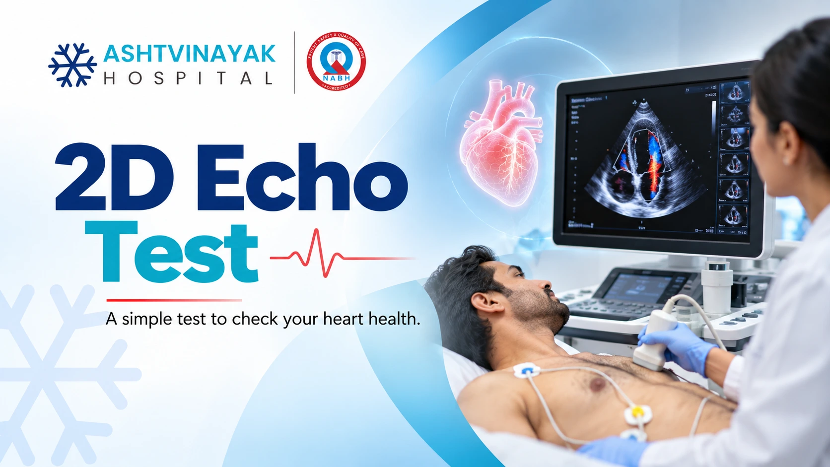


The heart continuously pumps blood throughout the body, and checking how well it works can be an important part of diagnosing cardiovascular problems. A 2D echo test is a commonly used heart imaging procedure that uses ultrasound waves to create moving images of the heart.
Unlike an X-ray or CT scan, echocardiography does not use ionising radiation. It can provide information about the size and structure of the heart chambers, valve function, movement of the heart muscle and how effectively the heart pumps blood. The test is generally painless and is performed without surgery.
What Is a 2D Echo Test?
A 2D echocardiogram, also called two-dimensional echocardiography, is an ultrasound examination of the heart. A handheld device called a transducer sends high-frequency sound waves into the body. These waves create images that allow the heart to be viewed while it is beating.
The test can show the heart chambers, valves, walls and surrounding structures. It can also provide information about blood flow when combined with Doppler echocardiography.
Doctors may recommend this investigation when symptoms, medical history or examination findings suggest that more information about heart function is needed.
Why Is a 2D Echo Test Done?
A 2D echocardiogram can help evaluate several aspects of heart health. It may be recommended for symptoms such as unexplained breathlessness, chest discomfort, swelling in the legs, fatigue or an abnormal heart sound detected during a physical examination.
Checking Heart Pumping Function
One important purpose of an echocardiogram is assessing how effectively the heart pumps blood. The left ventricle is particularly important because it pumps oxygen-rich blood to the rest of the body.
The test can help estimate the ejection fraction, which describes the percentage of blood pumped out of the left ventricle with each heartbeat.
Assessing Heart Valves
The heart has four valves that help blood move in the correct direction. Echocardiography can show whether valves are opening and closing properly and can help identify conditions such as valve narrowing or leakage.
Examining Heart Chambers
The test can provide information about the size and structure of the heart chambers. Enlargement of a chamber may occur with certain cardiovascular conditions and can be assessed through imaging.
Identifying Structural Problems
Echocardiography can help detect certain structural abnormalities affecting the heart muscle, walls or other cardiac structures. It can also be used to monitor a known heart condition over time.
How Is a 2D Echo Test Performed?
The procedure is usually performed while the patient is lying on an examination bed. Small adhesive electrodes may be placed on the chest to monitor the heart rhythm during the examination.
A healthcare professional applies a small amount of ultrasound gel to the chest and moves the transducer over different areas. The transducer sends sound waves into the body and receives the returning signals, which are converted into images on a monitor.
The patient may be asked to change position or briefly hold their breath so that clearer images can be obtained. The procedure is generally comfortable and does not involve needles or incisions.
How Long Does a 2D Echo Take?
The duration can vary depending on the type of echocardiogram and the amount of information required. A standard examination commonly takes around 30 to 60 minutes, although the actual time can differ between patients.
Once the images have been obtained, they are reviewed and interpreted by an appropriately qualified healthcare professional. The final report is considered alongside symptoms, medical history and other clinical findings.
Is Preparation Required Before a 2D Echo?
For a standard transthoracic echocardiogram, extensive preparation is generally not required. Patients can usually eat, drink and take their regular medicines unless their healthcare provider gives different instructions.
If a specialised type of echocardiogram is planned, preparation requirements may be different. Patients should follow the specific instructions provided by the healthcare facility.
Is a 2D Echo Test Painful?
A standard 2D echocardiogram is generally painless. Some pressure may be applied with the transducer while obtaining images from different parts of the chest, but it should not normally cause significant discomfort.
The ultrasound itself cannot be felt. Because the procedure does not involve radiation, injections or surgical incisions, it is commonly used for repeated assessment when clinically required.
What Conditions Can a 2D Echo Help Detect?
A 2D echocardiogram can provide useful information in several heart conditions. It may help assess heart failure, valve disorders, cardiomyopathy and certain congenital heart abnormalities.
It can also help evaluate changes in heart structure following certain cardiovascular events and monitor known heart conditions over time.
However, no single test can diagnose every heart problem. Depending on the symptoms, a doctor may also recommend an ECG, blood tests, stress testing, CT, MRI or other investigations.
Book your cardiac evaluation at Ashtavinayak Hospital for timely and appropriate heart care.
What Is Ejection Fraction in a 2D Echo?
Ejection fraction is a measurement used to assess the pumping function of the heart’s left ventricle. It represents the percentage of blood pumped out of the ventricle with each contraction.
A reduced ejection fraction can occur in certain forms of heart failure and other heart conditions. However, ejection fraction is only one part of a complete cardiac assessment. A doctor considers it alongside symptoms, examination findings and other test results.
What Happens After a 2D Echo?
A patient can generally return to normal activities immediately after a standard echocardiogram unless another procedure or medical condition requires different instructions.
The images are reviewed and a report is prepared. If the test was ordered because of specific symptoms, the doctor may discuss the results along with any additional investigations or treatment that may be needed.
An abnormal result does not necessarily indicate a severe condition. Its significance depends on the specific finding and the patient’s overall clinical picture.
When Should You Consider a Heart Check-Up?
Symptoms such as persistent chest discomfort, unexplained breathlessness, fainting, palpitations, unusual fatigue or swelling in the legs can have several causes. Some may be related to the heart and require medical evaluation.
People with conditions such as high blood pressure, diabetes or a known heart problem may also require periodic cardiovascular assessment according to their doctor’s recommendations.
Severe or sudden chest pain, significant difficulty breathing, fainting or other acute symptoms require urgent medical attention rather than waiting for a routine test.
Why Choose Ashtavinayak Hospital for a 2D Echo Test?
Ashtavinayak Hospital provides medical evaluation and diagnostic care for a range of health conditions. A cardiac investigation such as echocardiography can be useful when symptoms or clinical findings require assessment of heart structure and function.
The healthcare team can review the patient’s symptoms and medical history and recommend appropriate investigations based on the clinical situation. Test results can then be considered alongside other findings to guide further care.
Conclusion
A 2D echo test is a non-invasive cardiac imaging procedure that uses ultrasound to assess the heart’s structure and function. It can provide useful information about chambers, valves, heart muscle movement and pumping ability.
The test is generally comfortable and does not use radiation. If symptoms or medical history indicate a need for cardiac evaluation, a doctor can determine whether echocardiography or another investigation is appropriate.
FAQ
What is a 2D echo test used for?
A 2D echocardiogram is used to assess the structure and function of the heart. It can provide information about heart chambers, valves, heart muscle movement and pumping function.
Is a 2D echo test painful?
A standard echocardiogram is generally painless. The healthcare professional moves an ultrasound transducer over the chest, sometimes applying mild pressure to obtain clear images.
Does a 2D echo use radiation?
No. A standard echocardiogram uses high-frequency sound waves rather than ionising radiation.
How long does a 2D echo take?
A standard examination may take around 30 to 60 minutes, although the duration can vary depending on the patient and the type of assessment required.
Can I eat before a 2D echo?
For a standard transthoracic echocardiogram, fasting is generally not required. However, preparation can differ for specialised echocardiography procedures, so patients should follow the instructions provided by their healthcare provider.